Standing Tall
How surgeons at Stanford Children’s Health are correcting scoliosis with a novel procedure
By WP Creative Group
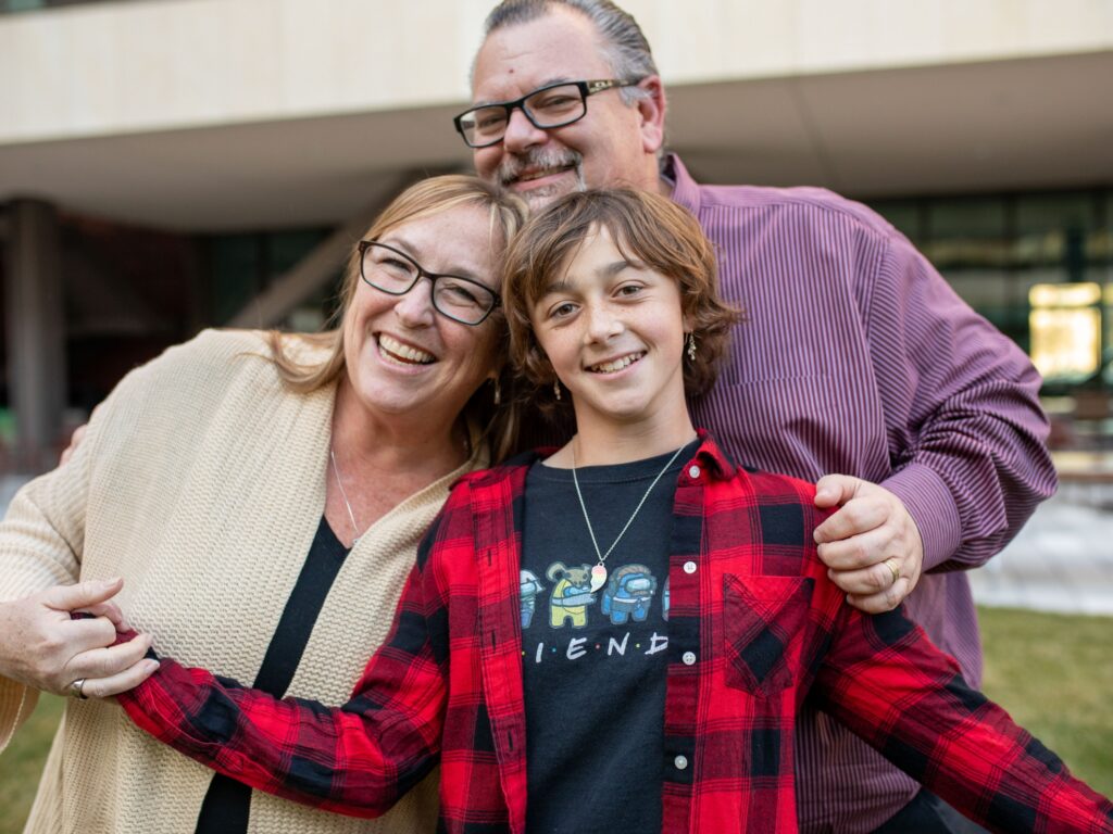
During a routine physical in 2019, Audrey Hersman’s pediatrician checked her spine and noticed a small lateral curve. This finding is relatively common, and Audrey had no symptoms, so her doctor planned to check again at her next visit. But thanks to the covid-19 pandemic, Audrey wouldn’t see her doctor again in person for more than a year. When she finally returned in 2021, it was clear: Audrey had severe scoliosis and would need surgery.
The news startled Audrey’s parents. Audrey still had no outward signs of a curve. Scoliosis affects 2% to 3% of Americans, but most patients do not meet the criteria for treatment. Surgery only becomes necessary when the curve reaches 45 degrees or more; Audrey’s curve was 50 degrees.
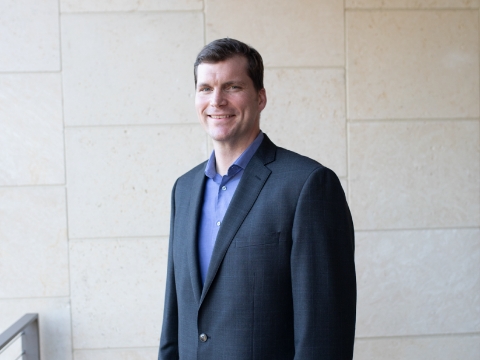
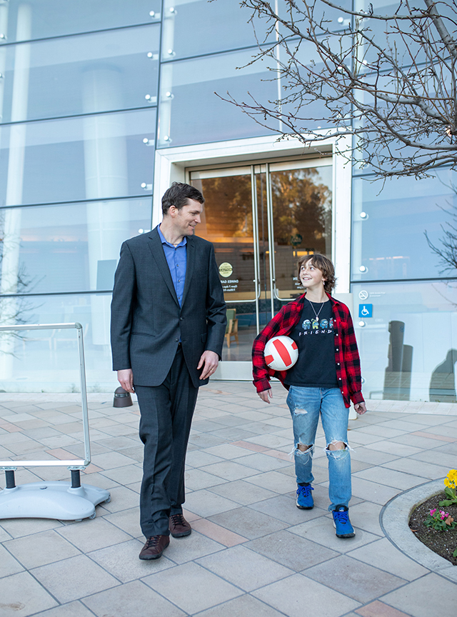
“Audrey is an example of a kid who wasn’t really having too many issues with her back, but over the years, her pediatrician started to notice that her back was asymmetrical,” said Dr. John Vorhies, a pediatric orthopedic surgeon at Stanford Children’s Health. “When it gets to the level that Audrey had, it will continue to worsen throughout growth and adulthood, and it can even affect the function of your lungs.”
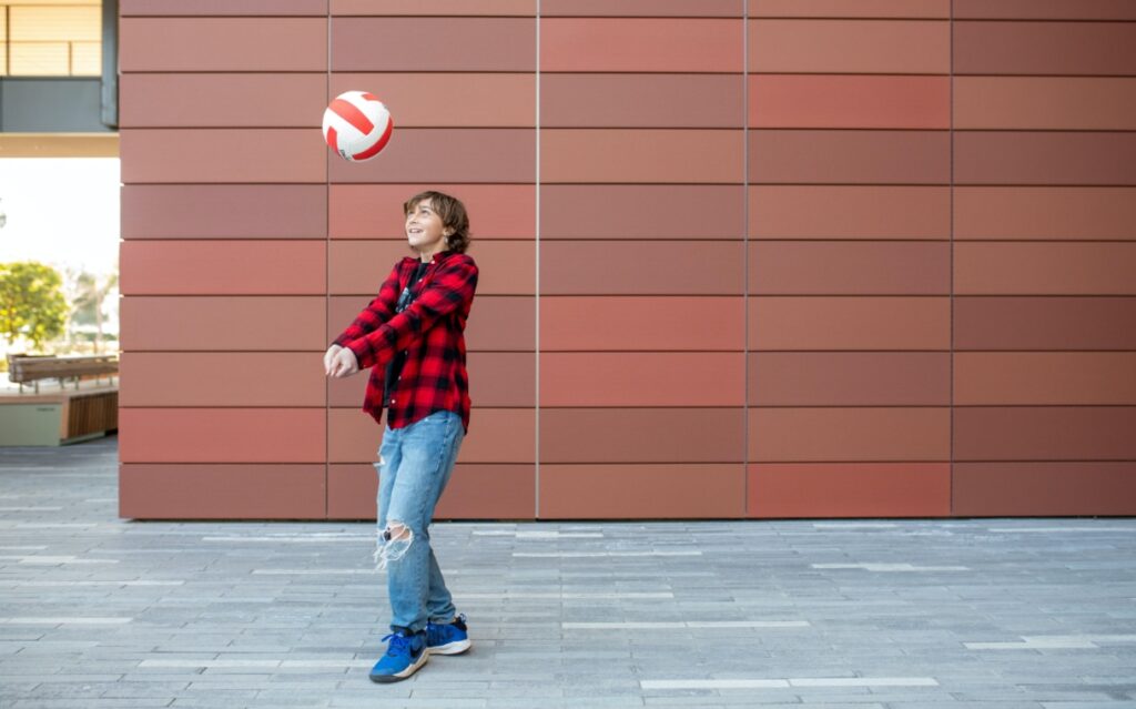
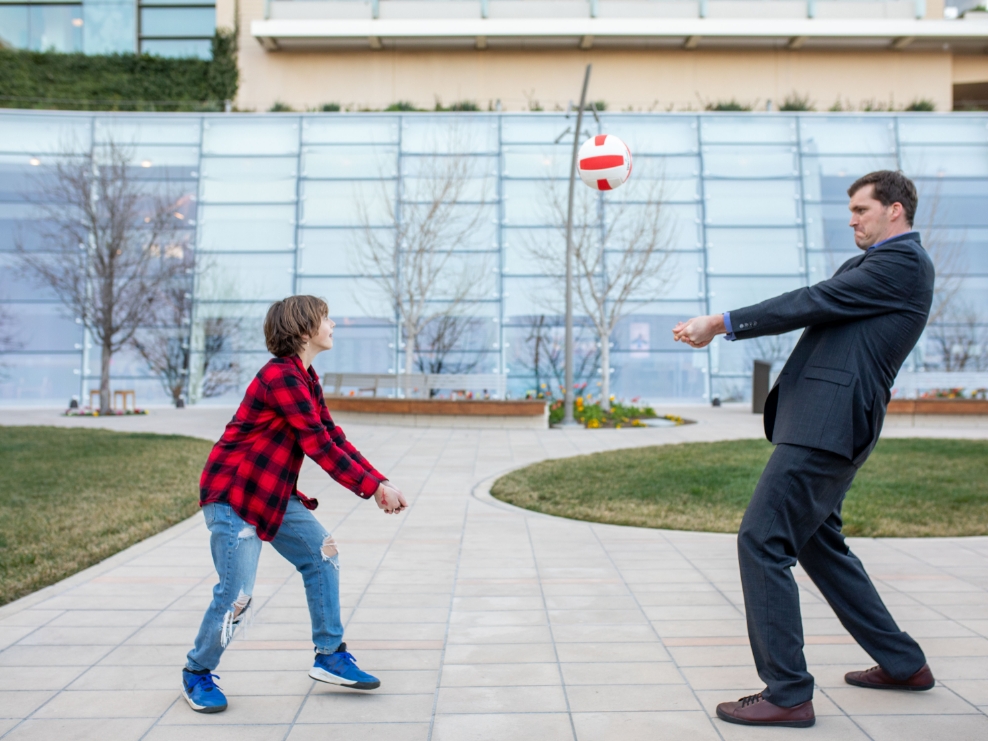
The traditional treatment for scoliosis is spinal fusion, in which surgeons straighten the spine by stabilizing vertebrae with screws and rods. Spinal fusion is a time-tested and highly effective procedure, but can leave lasting consequences, including limited mobility and higher risk of arthritis. The Hersmans wondered if there could be another option for Audrey, who was about to enter seventh grade and wanted to keep running track and playing volleyball.
“In many ways, Audrey was the ideal candidate for tethering. But my job is not just to come up with a treatment plan, but to really meet the family and the child where they are and come up with a targeted way to educate them about their options.“
-Dr. Vorhies
The Hersmans learned about a new kind of procedure: spinal tethering. It had only been approved by the U.S. Food and Drug Administration in 2019, but it had some potential benefits for a patient like Audrey. In spinal tethering, implants compress the outer side of the spinal curvature, allowing children’s natural growth to correct the curve as they age. It is a less invasive surgery than spinal fusion, places less pressure on the upper and lower spine and can maintain the back’s natural flexibility. And at Stanford Children’s Health, pediatric surgeons had already been training on how to do it.
Dr. Vorhies explained to the Hersman family that spinal tethering was still a new procedure, and the outcome was more uncertain than with spinal fusion. If it did not work as planned, Audrey might have to return for a spinal fusion. Audrey and her parents weighed the risks and decided that Audrey would be Stanford’s first spinal tethering patient.
From Audrey’s perspective, it was a chance to set an example.
I wanted to be a beacon for other kids, so they won’t be scared when they get the surgery…I wanted to be the first, so that there could be others to follow.
Audrey Hersman
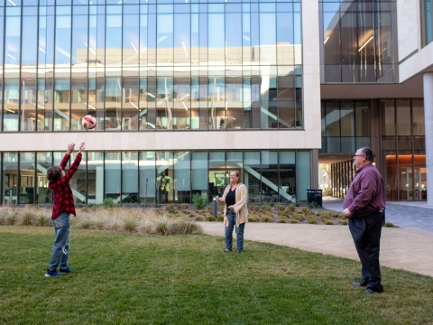
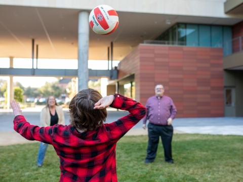
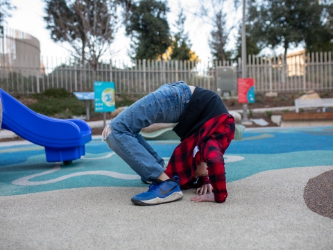
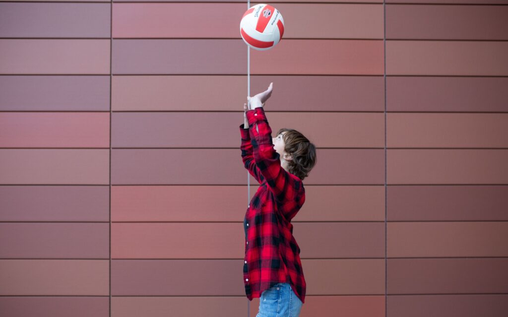
It was also an opportunity for her to keep playing volleyball with friends and continue to move freely as her spine continued to grow.
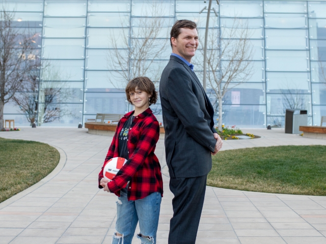
“She did a backbend for me in the clinic,” Dr. Vorhies said. “And she told me, ‘I want to be able to do that afterwards.’”
Pioneering a New Spinal Procedure
On the day of the surgery, while her parents waited outside, Audrey underwent a meticulous procedure that her surgeons had spent months preparing for.
To learn about spinal tethering, Dr. Vorhies and Dr. Matias Bruzoni had visited other hospitals to see how they practiced the technique, watched videos of the procedure together and consulted with representatives for the tools they planned to use. At the Mayo Clinic, Dr. Vorhies saw how surgeons used an O-arm, a 3-D imaging system, to navigate the spine more efficiently and safely. The team at Stanford Children’s adapted this idea to a different system called the Z-arm, which can take both 2-D and 3-D images.
Once they had the plan and the equipment, the team simulated the procedure on a mannequin in a Stanford Children’s operating room, holding a dress rehearsal before operating on Audrey.
“Pediatric surgery is a specialty where the surprise factor is a very common issue, and there’s always some level of anxiety when you do something for the first time,” said Dr. Bruzoni, a pediatric surgeon at Stanford Children’s Health. “The nice thing about collaboration in our group is that when we come across something we’ve never seen before, we call our partners and try to help each other.”
After nearly a year of preparation, Audrey underwent the tethering procedure last fall.
To begin the surgery, Dr. Bruzoni made a few five-millimeter incisions, about the width of a pencil eraser, in Audrey’s chest. Next, he used tiny tools to expose the spine and divide small blood vessels in the operative field so that Dr. Vorhies could access the vertebral bodies — the bones forming the front of each vertebra of her spine.
Typically, a procedure like this would require a very big incision that, for a child, often leads to significant deformities in the chest wall later in life.
“Children are still developing their chest wall and muscles, so the healing process is different than for adults, and it’s very important to minimize collateral damage,” Dr. Bruzoni said. “This technique results in a very good cosmetic outcome, where you can barely see the scars.”
Using a tiny camera and long instruments only five millimeters wide, the surgeons placed screws in Audrey’s vertebrae and attached the tethering implants, carefully avoiding the spinal cord and fragile bundles of blood vessels connecting the aorta to the chest wall.
We have a video-game-type array in the operating room, and we can see multiple three-dimensional images of the child’s spine that allow us to watch the implant go in safely
Dr. Vorhies
Standing Tall
Outside in the waiting room, the Hersmans got some good news: the surgery had been a success.
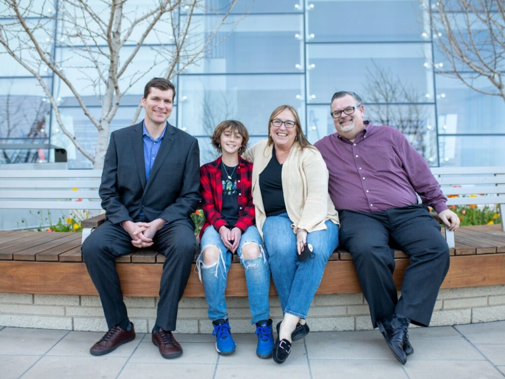
“The day of the surgery was really tough,” said Mark Hersman. “But it went perfectly. They took their time, and they were very meticulous about it.”
Audrey went home and recuperated after her time in the hospital and was back in school after two weeks. At her six-week follow-up, the news was better than she and her family could have hoped: Audrey had a clean bill of spine health, she didn’t even need to wear the night brace she had been prepared for, and she could return to playing sports.
Even better: with a straightened spine, Audrey grew almost two inches, which helps her on the volleyball court.
“So far, her treatment has been a success,” Dr. Vorhies said. “One of the reasons we were able to do it safely and have a good result was the multidisciplinary team we were able to mobilize, and the exhaustive amount of preparation and planning to make sure that there were no unknowns.”
The team included general surgeons, representatives from the implant company, surgical technicians and operating room nurses, all of whom participated in the staging event beforehand and then came together to fix Audrey’s spine in Stanford Children’s first spinal tethering surgery.
Audrey was expecting major scarring from the surgery, but she came away with just a few puncture marks on her side, where the laparoscopic tools entered her body. For Halloween, Amy Hersman made her daughter a skeleton costume with screws in the spine so Audrey could show off her new spinal accessories. They shared a photo with Audrey’s surgeons — proof of a successful surgery as well as the friendly, collaborative nature of the relationship between the family and their medical team.
It was nerve-wracking, but I’m really glad we did it…It has changed my life for the better.
Audrey Hersman
Learn more about Stanford Children’s Health Children’s Orthopedic and Sports Medicine Center.
Credits: By WP Creative Group